CIRSE 2014: Transbrachial approach for uterine leiomyoma embolization
Stefano PIERI, Paolo AGRESTI, Barbara SESSA, Maria Grazia Buquicchio, Vittorio MIELE
Purpose
To investigate the technical characteristic (feasibility, safety, X-ray exposure, side effects) of the transbrachial approach in uterine artery embolization.
Methods and materials
Between 2009 and 2013, 115 patients were treated with embolization of the uterine arteries for one or more symptomatic leiomyomas. In 20 of these 115 patients a transbrachial approach was used.
The inclusion criteria for all patients were the presence of one or more symptomatic fibromas, the desire to preserve fertility, the mainly intramural localization of the lesions, and the feasibility of embolization, determined by magnetic resonance imaging.
The transbrachial approach was carried out under ultrasound guidance, through the left brachial artery.
After having negotiated the entry of the descending thoracic artery [Fig. 1], the tip of the angiographic catheter, multipurpose 4 FR, was placed at the level of L4 to check the aortic bifurcation and subsequently, the uterine arteries were catheterized easily [Fig. 2]
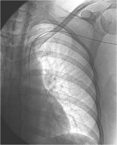 Fig . 1 – The guide wire is easily entered in the descending thoracic aorta, only turning the tip of the wire
The embolization was achieved with an injection of calibrated particles (500 and 700 microns).
Data concerning exposure to radiation and the duration of the intervention were recorded.
Clinical controls and MRI imaging were complemented with echo-colour Doppler of the brachial artery to confirm the integrity and function of the vessel.
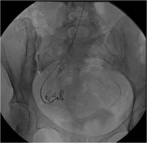 Fig. 2 – Selective catheterization of the right uterine artery only with the 4 Fr multipurpose, related to the hypertrophy of the artery.
Results
Uterine artery catheterization was easily performed in all patients (100%) with the multipurpose catheter, and the catheterization time for this group was less than that for the femoral group: 1,4’ was the median time for each uterine artery catheterization as compared with 3,4’ for the femoral group.
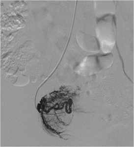 Fig. 3 – Visualization of the leiomyoma before embolization with hydophilic calibrated oarticles 500 e 700 micron
X-ray exposure was 21’ for the former group and 36’ in the latter one.
No immediate complications involving the brachial artery were recorded and in the seven patients who underwent echo-colour Doppler follow-up 1 year after the endovascular intervention, no changes were found in the morphology or blood flow in the vessels.
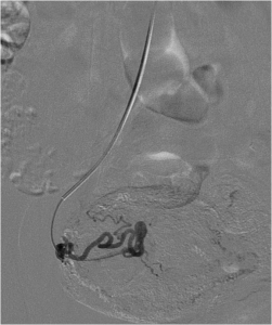 Fig. 4 -Visualization of the angiographic control at the end of the procedure.
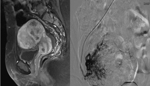 Leiomyoma before the procedure [Fig A] and during the angiographic control before right uterine artey embolization [Fig B] with hydrophilic calibrated particles 500 and 700 micron.
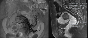 Leiomyoma during left uterine artery catheterization [Fig C], embolized only with hydrophilic calibrated particles 700 micron, for the visualization of the ovarian artery and RM control after three months [Fig D].
Conclusions
Transbrachial approach for uterine artery embolization is technically feasible, it is quick and easier than femoral approach; it permits deambulation after 2 or 3 hours from the procedure, easier observation of the percutaneous access and hospitalization for fewer days.